Review Sheet the Autonomic Nervous System Exercise 20
Affiliate 16. The Nervous System
16.4 The Peripheral Nervous Organization
Learning Objectives
By the end of this section, you will be able to:
- Describe the organization and functions of the sympathetic and parasympathetic nervous systems
- Describe the organization and part of the sensory-somatic nervous system
The peripheral nervous organisation (PNS) is the connectedness between the central nervous system and the rest of the body. The CNS is like the power plant of the nervous system. It creates the signals that control the functions of the trunk. The PNS is similar the wires that go to individual houses. Without those "wires," the signals produced past the CNS could not control the body (and the CNS would not be able to receive sensory data from the body either).
The PNS can be broken down into the autonomic nervous arrangement, which controls bodily functions without conscious control, and the sensory-somatic nervous system, which transmits sensory information from the skin, muscles, and sensory organs to the CNS and sends motor commands from the CNS to the muscles.
Autonomic Nervous System
Which of the post-obit statements is false?
- The parasympathetic pathway is responsible for resting the body, while the sympathetic pathway is responsible for preparing for an emergency.
- Nigh preganglionic neurons in the sympathetic pathway originate in the spinal cord.
- Slowing of the heartbeat is a parasympathetic response.
- Parasympathetic neurons are responsible for releasing norepinephrine on the target organ, while sympathetic neurons are responsible for releasing acetylcholine.
The autonomic nervous arrangement serves every bit the relay between the CNS and the internal organs. It controls the lungs, the heart, smooth muscle, and exocrine and endocrine glands. The autonomic nervous system controls these organs largely without conscious command; information technology can continuously monitor the conditions of these different systems and implement changes equally needed. Signaling to the target tissue commonly involves ii synapses: a preganglionic neuron (originating in the CNS) synapses to a neuron in a ganglion that, in turn, synapses on the target organ, as illustrated in Figure 16.26. There are 2 divisions of the autonomic nervous organization that frequently accept opposing furnishings: the sympathetic nervous system and the parasympathetic nervous system.
Sympathetic Nervous Organization
The sympathetic nervous system is responsible for the "fight or flight" response that occurs when an animal encounters a dangerous situation. One way to remember this is to think of the surprise a person feels when encountering a ophidian ("snake" and "sympathetic" both begin with "due south"). Examples of functions controlled by the sympathetic nervous system include an accelerated eye rate and inhibited digestion. These functions help prepare an organism's body for the physical strain required to escape a potentially unsafe situation or to fend off a predator.
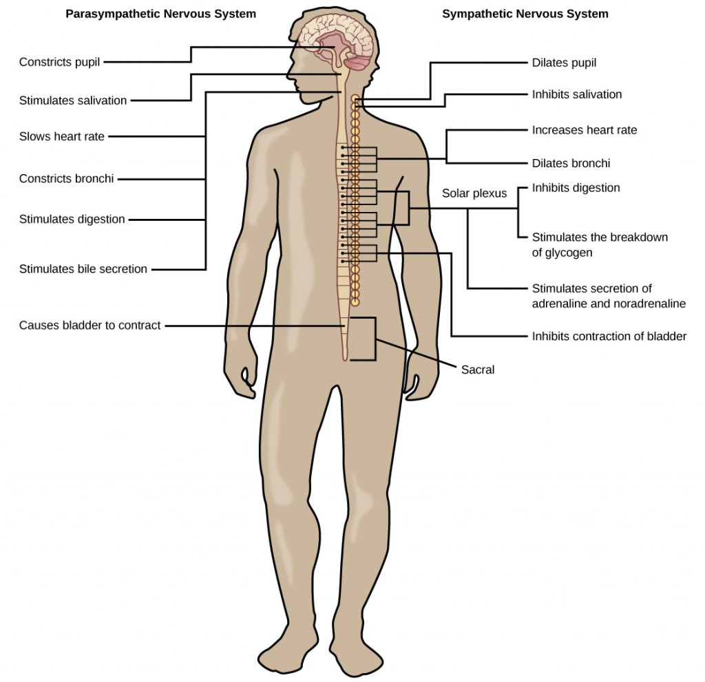
Most preganglionic neurons in the sympathetic nervous organization originate in the spinal cord, as illustrated in Figure 16.27. The axons of these neurons release acetylcholine on postganglionic neurons within sympathetic ganglia (the sympathetic ganglia class a concatenation that extends alongside the spinal cord). The acetylcholine activates the postganglionic neurons. Postganglionic neurons and then release norepinephrine onto target organs. As anyone who has ever felt a rush before a big test, speech, or athletic event can attest, the furnishings of the sympathetic nervous system are quite pervasive. This is both considering one preganglionic neuron synapses on multiple postganglionic neurons, amplifying the effect of the original synapse, and considering the adrenal gland also releases norepinephrine (and the closely related hormone epinephrine) into the claret stream. The physiological effects of this norepinephrine release include dilating the trachea and bronchi (making it easier for the animal to breathe), increasing heart rate, and moving claret from the peel to the centre, muscles, and encephalon (then the animal can think and run). The strength and speed of the sympathetic response helps an organism avoid danger, and scientists accept found evidence that it may also increment LTP—allowing the animal to think the dangerous state of affairs and avoid it in the future.
Parasympathetic Nervous Organization
While the sympathetic nervous system is activated in stressful situations, the parasympathetic nervous system allows an animal to "rest and digest." One fashion to remember this is to call back that during a restful situation like a picnic, the parasympathetic nervous system is in control ("picnic" and "parasympathetic" both kickoff with "p"). Parasympathetic preganglionic neurons have cell bodies located in the brainstem and in the sacral (toward the bottom) spinal cord, as shown in Figure 16.27. The axons of the preganglionic neurons release acetylcholine on the postganglionic neurons, which are generally located very near the target organs. Nearly postganglionic neurons release acetylcholine onto target organs, although some release nitric oxide.
The parasympathetic nervous system resets organ function later on the sympathetic nervous system is activated (the mutual adrenaline dump you feel after a 'fight-or-flight' event). Furnishings of acetylcholine release on target organs include slowing of center rate, lowered blood pressure, and stimulation of digestion.
Sensory-Somatic Nervous Arrangement
The sensory-somatic nervous organization is made upwardly of cranial and spinal nerves and contains both sensory and motor neurons. Sensory neurons transmit sensory information from the skin, skeletal musculus, and sensory organs to the CNS. Motor neurons transmit messages near desired motion from the CNS to the muscles to make them contract. Without its sensory-somatic nervous organisation, an animal would exist unable to process any information about its environment (what it sees, feels, hears, and then on) and could not control motor movements. Unlike the autonomic nervous organisation, which has two synapses between the CNS and the target organ, sensory and motor neurons accept just one synapse—one catastrophe of the neuron is at the organ and the other directly contacts a CNS neuron. Acetylcholine is the main neurotransmitter released at these synapses.
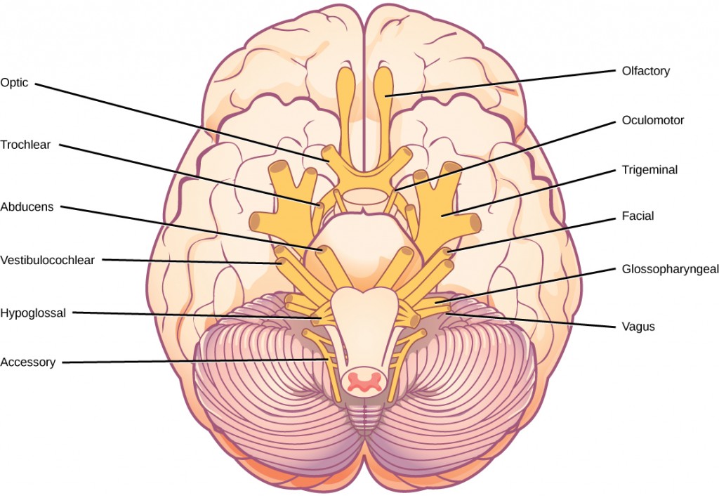
Humans accept 12 cranial nerves, nerves that sally from or enter the skull (cranium), every bit opposed to the spinal nerves, which emerge from the vertebral column. Each cranial nervus is accorded a proper name, which are detailed in Figure 16.28. Some cranial nerves transmit only sensory data. For case, the olfactory nervus transmits data about smells from the nose to the brainstem. Other cranial nerves transmit almost solely motor information. For case, the oculomotor nerve controls the opening and closing of the eyelid and some eye movements. Other cranial nerves contain a mix of sensory and motor fibers. For instance, the glossopharyngeal nerve has a role in both taste (sensory) and swallowing (motor).
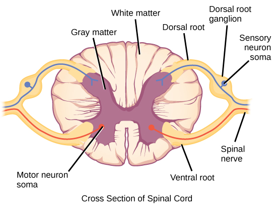
Spinal fretfulness transmit sensory and motor information between the spinal cord and the rest of the torso. Each of the 31 spinal nerves (in humans) contains both sensory and motor axons. The sensory neuron jail cell bodies are grouped in structures called dorsal root ganglia and are shown in Effigy sixteen.29. Each sensory neuron has one project—with a sensory receptor ending in skin, muscle, or sensory organs—and another that synapses with a neuron in the dorsal spinal cord. Motor neurons have jail cell bodies in the ventral gray affair of the spinal cord that project to musculus through the ventral root. These neurons are usually stimulated by interneurons within the spinal cord merely are sometimes directly stimulated by sensory neurons.
Summary
The peripheral nervous system contains both the autonomic and sensory-somatic nervous systems. The autonomic nervous organisation provides unconscious control over visceral functions and has 2 divisions: the sympathetic and parasympathetic nervous systems. The sympathetic nervous arrangement is activated in stressful situations to fix the animal for a "fight or flying" response. The parasympathetic nervous system is active during restful periods. The sensory-somatic nervous system is fabricated of cranial and spinal nerves that transmit sensory information from peel and musculus to the CNS and motor commands from the CNS to the muscles.
Exercises
- Which of the post-obit statements is false?
- The parasympathetic pathway is responsible for relaxing the body, while the sympathetic pathway is responsible for preparing for an emergency.
- Most preganglionic neurons in the sympathetic pathway originate in the spinal cord.
- Slowing of the heartbeat is a parasympathetic response.
- Parasympathetic neurons are responsible for releasing norepinephrine on the target organ, while sympathetic neurons are responsible for releasing acetylcholine.
- Activation of the sympathetic nervous system causes:
- increased blood flow into the skin
- a decreased heart charge per unit
- an increased center charge per unit
- increased digestion
- Where are parasympathetic preganglionic cell bodies located?
- cerebellum
- brainstem
- dorsal root ganglia
- skin
- ________ is released by motor nerve endings onto muscle.
- Acetylcholine
- Norepinephrine
- Dopamine
- Serotonin
- What are the main differences between the sympathetic and parasympathetic branches of the autonomic nervous organization?
- What are the main functions of the sensory-somatic nervous system?
Answers
- D
- C
- B
- A
- The sympathetic nervous arrangement prepares the trunk for "fight or flight," whereas the parasympathetic nervous arrangement allows the trunk to "rest and digest." Sympathetic neurons release norepinephrine onto target organs; parasympathetic neurons release acetylcholine. Sympathetic neuron cell bodies are located in sympathetic ganglia. Parasympathetic neuron prison cell bodies are located in the brainstem and sacral spinal cord. Activation of the sympathetic nervous system increases heart rate and blood force per unit area and decreases digestion and claret flow to the peel. Activation of the parasympathetic nervous system decreases heart rate and blood pressure and increases digestion and blood flow to the skin.
- The sensory-somatic nervous system transmits sensory information from the skin, muscles, and sensory organs to the CNS. Information technology likewise sends motor commands from the CNS to the muscles, causing them to contract.
Glossary
- acetylcholine
- neurotransmitter released past neurons in the key nervous system and peripheral nervous system
- autonomic nervous arrangement
- part of the peripheral nervous organization that controls bodily functions
- cranial nerve
- sensory and/or motor nerve that emanates from the brain
- norepinephrine
- neurotransmitter and hormone released by activation of the sympathetic nervous system
- parasympathetic nervous system
- division of autonomic nervous system that regulates visceral functions during balance and digestion
- sensory-somatic nervous system
- system of sensory and motor fretfulness
- spinal nerve
- nerve projecting between peel or muscle and spinal cord
- sympathetic nervous system
- division of autonomic nervous system activated during stressful "fight or flight" situations
Source: https://opentextbc.ca/biology/chapter/16-4-the-peripheral-nervous-system/
0 Response to "Review Sheet the Autonomic Nervous System Exercise 20"
Post a Comment